Osteoarthritis of the knee, an increasingly common health problem among active adults, is a painful condition involving loss of cartilage and deformity of the joint. It is also a major factor in diminished mobility, especially among older people.
Multiple studies have identified stress injuries to the leg bones as a primary contributor to arthritic knee pain and deformity. Little is known, however, about the causes and progression of bone-stress injuries among middle-aged, active adults.
Kai-Yu Ho, an assistant professor in the School of Allied Health Sciences, is directing a study that could remedy this deficiency, while at the same time providing a method for evaluating the probability that osteoarthritis is present. She also hopes to help doctors detect the sort of bone damage that produces osteoarthritis, even before a person experiences pain.
Ho's study involves an examination of how "locomotion-induced shock loading" -- the results of such everyday, bone-jarring movement like walking, running, and stair climbing -- affects the physiology of knees. Her work also examines the alignment of bones in legs that have stress injuries, and how injury-related alignment issues might relate to arthritis development.
A normal femur, or upper leg bone, is not perfectly vertical. The bone extends from the hip at a slight angle toward the midline of the body. Normal physical activity can cause micro-damage to the femur and tibia where they meet at the knee. The body repairs the damage by growing new bone, a process called bone remodeling.
Continual shock-loading damage can cause too much bone remodeling, however, with new bone forcing the femur away from the midline of the body into a "varus angle" (a phrase adapted from the Latin word for "crooked"). When the varus angle is pronounced a person appears bowlegged -- the knees do not touch when one stands with his or her feet together.
Ho hypothesizes that increased shock loading among middle-aged persons will lead to greater bone stress, which can increase the onset of osteoarthritis in the joint. She put her hypothesis to the test in a study involving 20 adults ranging in age from 50 to 65. None of her subjects had been diagnosed with arthritis in the knee.
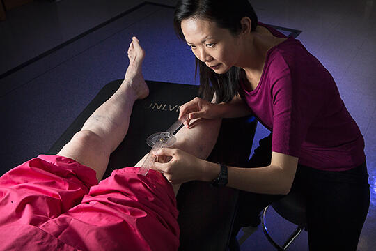
Participants were asked to complete a locomotion-induced, shock-loading activity. When finished, they receive a magnetic resonance imaging (MRI) scan to verify bone stress injuries. Participants next allowed the researchers to measure the angle between the long axis of their femurs and tibias.
Ho says she and her team are now analyzing the data they've collected to determine whether there is an association between lower extremity alignment and MRI-detected bone stress injury. If there is, she says, the finding could provide health-care providers with an early detection method of bone abnormalities. This, in turn, will enable them to recommend physical therapy intervention to ward off future bone-stress injuries to patients' knees.
Research related to this study has been published in the European Journal of Sports Science and Magnetic Resonance Imaging.


